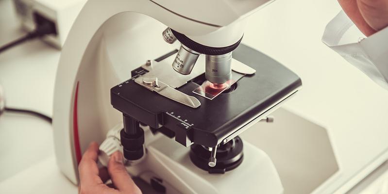
 Histology, regularly called the cornerstone of cutting-edge remedies, delves into the tough global of cell shape, revealing the microscopic wonders that underpin the functioning of tissues and organs. This problem of having a look at, moreover called microscopic anatomy, is pivotal in unraveling the complexities of natural structures, supplying treasured insights into the inner workings of dwelling organisms. At the coronary heart of histology lies the meticulous examination of tissue samples, a manner that unveils the hidden landscapes of cells and their complicated interactions in the frame.
These specialized centers are the epicenters of discovery, wherein dedicated histology technicians, armed with precision gadgets and unwavering understanding, resolve the mysteries of cellular morphology and commercial enterprise business enterprise. Through a symphony of staining strategies, tissue embedding strategies, and sectioning methodologies, histology labs rework ordinary tissue samples into home windows into the microscopic universe, supplying researchers and pathologists top-notch insights into the cloth of lifestyles itself.
Role of Histology Labs
Histology labs are integral pillars within the nation-states of medical analysis, pathology, and organic research, serving as the gatekeepers to the microscopic realm of tissues and cells. At the helm of those centers are histology technicians, professional specialists whose information lies inside the problematic handling and processing of tissue samples. Their meticulous work includes a spectrum of responsibilities, starting from the preliminary receipt of tissue specimens to the very last instruction of slides for microscopic examination.
Sample Processing and Preparation:
Histotechnologists are tasked with the sensitive tissue processing system, a critical step that ensures the upkeep of tissue structure and mobile morphology. This manner entails a series of meticulously orchestrated steps, inclusive of fixation, dehydration, clearing, and infiltration with an embedding medium. Through those steps, histology labs rework fragile tissue samples into sturdy specimens appropriate for similar evaluation below the microscope.
Microscopic Analysis:
Once tissue samples are appropriately processed and embedded, they go through sectioning—a procedure wherein skinny slices of tissue, referred to as histological sections, meticulously reduce the usage of specialized devices called microtomes. These sections are then set up on glass slides and subjected to numerous staining strategies to beautify cell structures and spotlight precise components of interest. Histotechnologists hire a diverse array of staining strategies, starting from ordinary hematoxylin and eosin staining to specialized stains like periodic acid-Schiff and immunohistochemistry, each offering precise insights into tissue composition and pathology.
Specialized Staining Techniques:
Histology labs are renowned for his or her skill in using specialized staining strategies to find elaborate information within tissue samples. Immunohistochemistry, as an instance, utilizes antibodies to come across particular proteins inside tissues, enabling pathologists to diagnose diverse illnesses and characterize tumor markers. Similarly, special stains including Masson's trichrome and Alcian blue are employed to highlight collagen and mucin, respectively, helping in the prognosis of connective tissue problems and gastrointestinal illnesses.
In essence, histology labs function as the cornerstone of diagnostic remedy and biomedical studies, providing beneficial insights into the intricate tapestry of tissues and organs. Through their meticulous sample processing techniques, microscopic analyses, and specialized staining methodologies, these facilities empower healthcare professionals and researchers alike to get to the bottom of the mysteries of disease pathology and pave the way for novel therapeutic interventions.
Applications of Histology
Histology, with its roots deeply embedded within the microscopic exploration of tissues, extends its some distance-reaching applications throughout diverse domains, showcasing its significance in each clinical diagnosis and biological research. This intricate branch of technological know-how plays a pivotal position in unraveling the mysteries held inside the cellular tapestry, providing insights that span from ailment prognosis to fundamental organic exploration.
Medical Diagnosis and Pathology:
Histology stands as a necessary tool in the palms of medical experts, mainly pathologists, who wield its electricity to diagnose a myriad of sicknesses. In the area of medical diagnosis, histological analysis serves as a beacon, guiding clinicians via the identification of cell abnormalities, tissue adjustments, and the complicated signatures of numerous illnesses. From cancerous growths to infectious sellers and inflammatory situations, histology acts as a diagnostic compass, enabling accurate disease characterization and paving the manner for informed remedy selections.
The microscopic examination of tissue samples, more advantageous with the aid of various staining techniques, presents a detailed map of cell structures, permitting pathologists to parent abnormalities, examine disorder development, and provide unique prognoses. In most cancers prognosis, as an instance, histological analysis now not only identifies the presence of tumors however additionally classifies their kind, grade, and degree—critical records that form the idea of tailor-made remedy techniques.
Biological Research:
Histology's significance transcends the realm of medication, extending its impact into the expansive area of biological studies. Researchers in the nation-states of embryology, physiology, and pathology turn to histological techniques to get to the bottom of the intricacies of tissue structure and function. In organic research, histology serves as a window into the inner workings of residing organisms, offering essential insights that gasoline improvements in biomedical technology.
Histological research contributes to our understanding of tissue improvement, organ feature, and the underlying mechanisms of illnesses. In embryology, histology unveils the complicated methods of organogenesis and tissue differentiation, shedding mild on the early levels of existence. Physiological studies advantage from histological analyses, permitting researchers to discover the cellular basis of organ function and homeostasis. In pathology studies, histology serves as a fundamental tool for investigating ailment mechanisms, identifying potential healing objectives, and validating the efficacy of interventions.
Histology's packages are always-reaching and impactful, shaping each the analysis and treatment of illnesses within the medical realm and contributing foundational expertise to the ever-evolving landscape of biological research. Through its microscopic lens, histology continues to be a cornerstone in advancing our expertise of the complexities inherent in existence and disease.
 Techniques Used in Histology Labs
Histology labs are bastions of precision and meticulousness, employing an arsenal of specialized strategies to unencumbered the secrets held within tissue samples. From tissue fixation to staining, each step within the histological manner is cautiously orchestrated to maintain cell integrity and monitor the problematic architecture of tissues underneath the microscope.
Tissue Fixation and Embedding:
At the heart of histological education lies tissue fixation, a method important for maintaining cellular morphology and preventing degradation. Formalin, a commonly used fixative, pass-links proteins within the tissue, locking them in region and halting enzymatic activity. Once fixed, tissues are embedded in a solid medium, usually paraffin wax or resin, to offer assistance for the duration of sectioning. Embedding guarantees that tissue samples hold their structural integrity and helps the production of skinny, uniform sections for microscopic evaluation.
Sectioning and Staining:
With embedded tissues in hand, histotechnologists continue the sensitive artwork of sectioning, in which tissue blocks are sliced into thin sections using specialized gadgets called microtomes. These sections, frequently only a few micrometers thick, are then mounted onto glass slides, geared up to undergo staining. Staining strategies are a hallmark of histological analysis, improving the assessment between mobile components and enabling visualization under the microscope. Hematoxylin and eosin staining, a cornerstone of histology, imparts colour to cellular nuclei and cytoplasm, respectively, making an allowance for distinctive exam of tissue structure and cellular morphology.
Additional stains, which includes periodic acid-Schiff and immunohistochemical stains, provide further insights into precise cell systems and molecular markers, expanding the diagnostic competencies of histological evaluation.
The strategies employed in histology labs constitute a sensitive balance between art and science, where precision and attention to detail converge to reveal the hidden intricacies of lifestyles on the cellular level. Through those strategies, histologists unravel the mysteries held inside tissue samples, paving the way for more desirable knowledge and diagnosis of diseases.
LabLink's Support for Histology Labs
Histology labs, with their intricate strategies and specialized devices, depend on seamless operations to preserve efficiency and precision in their paintings. Recognizing the specific needs of these centers, LabLink gives tailor-made aid answers aimed toward enhancing productivity and making sure the highest standards of overall performance.
Equipment Management Solutions:
- LabLink is familiar with the critical function that gadget plays in histology labs and offers complete control solutions to deal with the unique desires of these facilities.
- From the procurement of current instrumentation to the disposal of old devices, LabLink publications histology labs via each degree of the equipment lifecycle.
- Expertise in equipment valuation permits histology labs to make knowledgeable selections concerning asset management, ensuring most beneficial aid allocation and budgetary performance.
- Additionally, LabLink's logistical assistance streamlines system transactions, facilitating seamless transitions and minimizing disruptions to lab operations.
Testimonials from Histology Lab Clients:
- Histology labs which have partnered with LabLink commend the corporation's willpower to handing over dependable offerings and exceeding expectations.
- Testimonials from glad clients highlight LabLink's professionalism, expertise, and commitment to client satisfaction.
- By entrusting their device management needs to LabLink, histology labs were capable of optimize their operations, reducing prices, and decorate performance.
- These testimonials function as a testimony to LabLink's reputation as a depended on associate for histology labs looking for green and powerful gadget management answers.
LabLink's aid for histology labs extends beyond mere service provision, encompassing a dedication to excellence and a willpower to assemble the precise desires of each facility. Through tailor-made solutions and unwavering reliability, LabLink empowers histology labs to recognize their central task of advancing scientific discovery and enhancing affected person care.
